TrazaScan 3D

60 seconds of video.
3D model.

A dental scanner captures thousands of photos and turns them into 3D. Your phone records 4K at 60fps — 3,600 frames per minute. TrazaScan 3D turns those into geometry.

60s
video capture
~120μm
accuracy
$0
extra equipment
The full process

From pixels to
geometry.

Pick your role. See your full flow — from capture to 3D model.

01
Surgeon recording dental video with phone
CHAIRSIDE
Record from TrazaScan

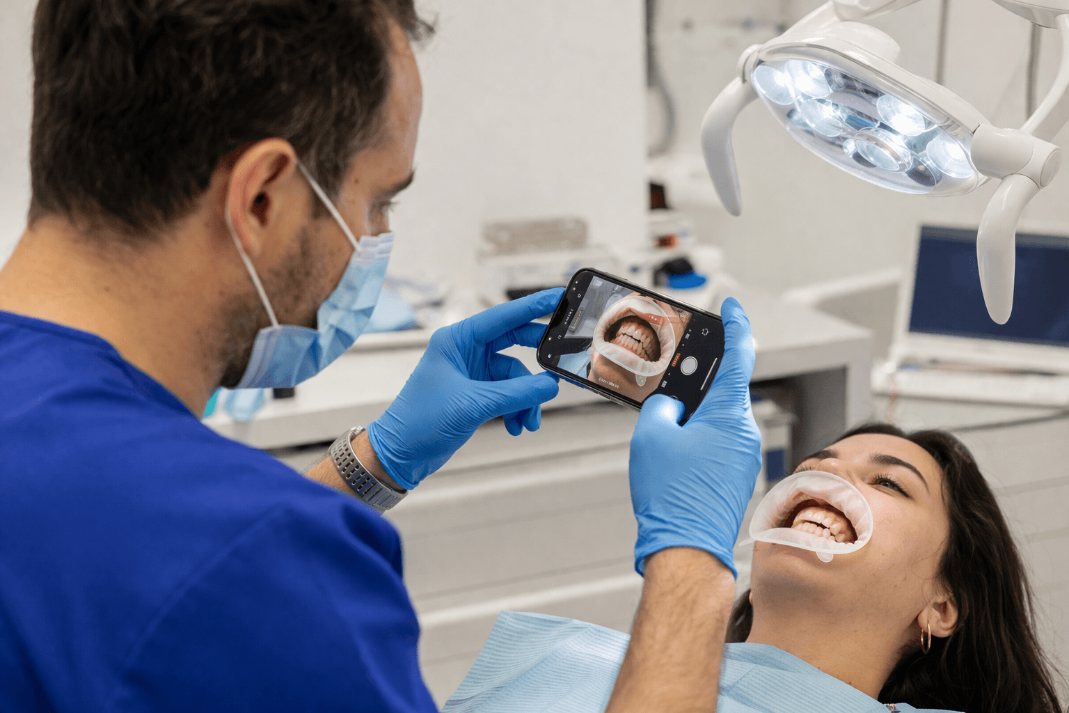
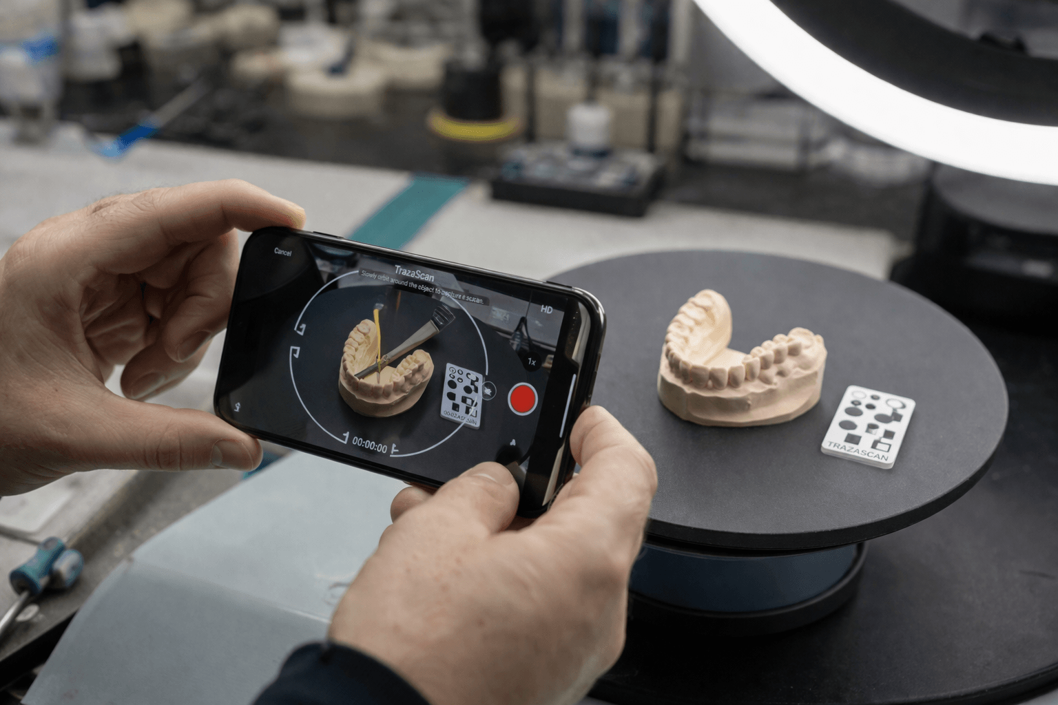
Open the tool. Record. Done.

From TrazaScan in your dashboard, turn on the camera and record 60 seconds orbiting the mouth with retractors. The visual guide tells you speed and coverage. When you finish, the video uploads to the platform automatically.
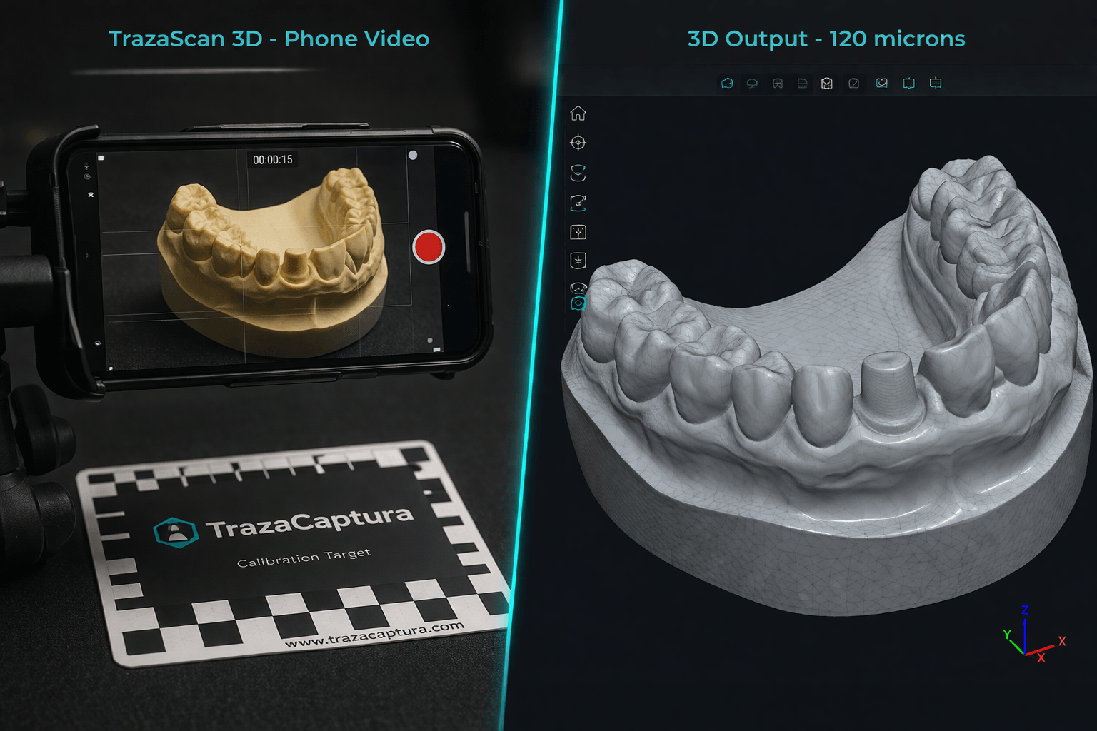
TrazaScan · 4K · 60fps · Auto upload
02
Automatic selection

~120 optimal frames

Out of thousands of frames, the system picks the best by sharpness, angular coverage, and consistent exposure. Redundant and blurry ones are dropped. Each frame is a high-resolution photo.
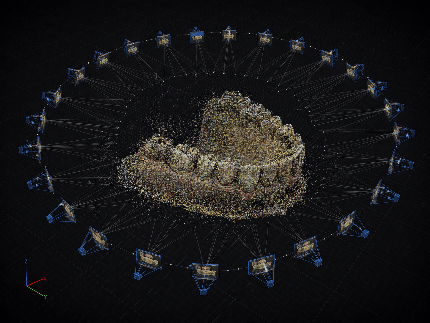
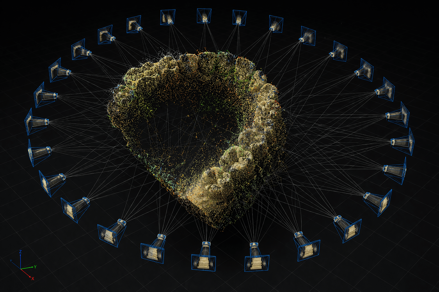
120 of 3,600 · Auto-filtered · HD
Frames extracted from dental video
EXTRACTION
03
SIFT feature detection on dental photo
MATCHING
SIFT + FLANN

Key points matched

Each frame is analyzed pixel by pixel. SIFT detects edges, cusps, margins. Each point gets a 128-dimension descriptor. They are matched across frames to triangulate 3D positions.

8,192 features · 128-dim · Matched
04
Patch-Match Stereo

Dense point cloud

Bundle Adjustment computes the 3D position of each camera. Then Patch-Match generates a dense depth map. Hundreds of thousands of 3D points — dental anatomy in three dimensions.
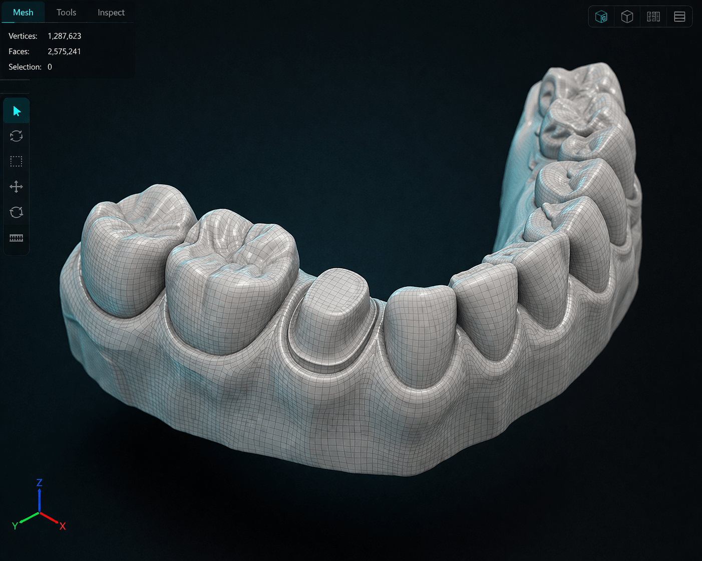
500K+ points · Depth · 3D
Dense point cloud of dental model
DENSE
3D
Reconstructed dental 3D model
RESULT
Poisson Surface Reconstruction

Final 3D model

Continuous mesh calibrated to real scale. Color texture extracted from the original images. Downloadable PLY, OBJ, or STL. Compatible with Exocad, 3Shape, Dental Wings.

~120μm · PLY/OBJ/STL · With texture

What you gain as a surgeon

Verify prep in 3D
Review the margin from every angle BEFORE taking the impression. Catch issues that direct view hides.
Preview for the lab
The lab sees your prep in 3D before the box arrives. Resolves doubts without a call.
3D evidence
Disputes resolved with geometry, not memory. Permanent record of what you sent.
Export STL/PLY
Compatible with any dental CAD. Share with any lab in the world.
The tech you already know

Thousands of photos. One mesh.

A dental scanner is not magic. Explore every step of the process.

Structured light
Projecting patterns onto the dental surface

The scanner projects stripes, dots, or light grids onto the tooth or model. The camera captures how those patterns deform when they hit the geometry. Each deformation encodes depth.

1-5Sensor megapixels
60+Captures per second
LEDControlled lighting
Depth data
Each deformed pattern = one fragment of a 3D map

The camera records how each pattern curves, stretches, or compresses. That deformation translates mathematically into depth data. Each frame produces a partial fragment of the model.

ZDepth map
Sub-pxSub-pixel accuracy
Real-timeInstant processing
ICP Registration
3,000-10,000 fragments fused into one continuous mesh

The software aligns thousands of partial 3D fragments using ICP (Iterative Closest Point). Each fragment is rotated and translated until points match. The result is a continuous surface with no gaps.

10KFused frames
ICPRegistration algorithm
0Gaps in the mesh
The cost of accuracy
STL mesh with 10-20μm — exceptional, but with barriers

Cost: $3,500 (desktop) to $40,000 (intraoral). Learning curve: 2-4 weeks. Annual calibration required. Exceptional result — but access barriers that shut out 51% of labs.

$3.5KMinimum price
10-20Microns of accuracy
AnnualCalibration required

The number that matters: A dental scanner sensor has 1-5 megapixels. Your iPhone has 48-200. The difference is not in the sensor — it is in the controlled lighting and the software.

The process

No scanner. No excuses. No limit.

Your video enters the COLMAP pipeline. Five stages of computational processing before it becomes 3D geometry.

Feature Detection

SIFT identifies unique points in each image: edges, corners, textures. Each point gets a 128-dimension descriptor that makes it unique and identifiable across frames.

Feature Matching

FLANN finds correspondences between descriptors in each image pair. Points that appear in multiple frames are linked to calculate their 3D position.

Sparse Reconstruction

Bundle Adjustment computes the 3D position of each camera (frame) and generates a sparse point cloud. It is the geometric skeleton of the model.

Dense Reconstruction

Patch-Match Stereo generates a dense depth map for each image. Millions of 3D points replace the sparse cloud. The anatomy takes shape.

Meshing

Poisson Surface Reconstruction turns the dense cloud into a continuous mesh (PLY/OBJ) with color texture extracted from the original images. 15-30 min on server.

No filters

The numbers nobody publishes.

We are not going to claim it replaces a $40,000 scanner. We are going to show exactly where each one stands.

Metric TrazaScan 3D Desktop Scanner Intraoral Scanner
Accuracy~80-150μm7-15μm10-20μm
Cost$0 (phone + TrazaLab)$3,500-7,000$25,000-45,000
Training5 minutes1-2 days2-4 weeks
Speed60s capture + 15-30min process2-5 min scan3-8 min scan
Output formatPLY / OBJSTLSTL
Mill from the model?Not yet (getting close)YesYes
Shade captureYes (TrazaTono built in)NoLimited
Clinical photosYes (bilateral)NoNo
Works offline?Capture yes, processing noYesYes
MaintenanceNoneAnnual calibrationAnnual calibration
Real use cases

120 microns is enough for this.

Accuracy that covers a broad set of applications where manufacturing tolerances are not required.

3D documentation

Every model that passes through the lab stays in 3D. Permanent archive without physical space.

Surgeon communication

A PLY shared in TrazaChat replaces 10 minutes of verbal description.

Orthodontics

Study, progress, and final models. All in 3D, no boxes. Automatic time-based comparison.

Planning

Show the patient their current model in 3D. Base for digital planning without a scanner.

Training

Technicians study real 3D models from any angle. Instructors annotate directly on the mesh.

Before/after comparison

Overlay 3D models from different stages. Quantify changes with direct measurements on the mesh.

Simple posteriors

Monolithic zirconia crowns: accuracy is getting close. With caveats.

Getting close
Transparency

Where we fall short.

Being honest about limits builds trust. Click each case.

Veneers, anterior crowns in lithium disilicate. Margins and surface texture need <50μm. TrazaScan sits at 80-150μm. Not enough for high-end esthetics.

Full arch demands cumulative accuracy over long distances. A 100μm error × 12 positions yields unacceptable passive misfit. Needs a dedicated scan.

CAD/CAM demands <25μm to mill without adjustment. TrazaScan produces meshes with 80-150μm of variability. The gap is real for most cases.

Photogrammetry needs direct line of sight. A margin 2mm subgingival has no visibility. Intraoral scanners also struggle here, but handle depth better.

Spaces between teeth are hard to capture with orbital video. The camera cannot access the angle needed to resolve interproximal geometry with enough precision.

Evolution

The gap closes every year.

Every year, phone cameras improve. Every year, reconstruction algorithms improve. The gap closes. TrazaScan 3D gets better automatically — no new hardware to buy.

Phase 1 — Now
Photographic documentation + extraction engine

Clinical photo capture with TrazaTono (color calibration), bilateral logging, and smart frame extraction from video. The base everything else is built on.

Phase 2 — Now
Video to 3D at ~120μm

Full COLMAP photogrammetry pipeline. From 4K video to PLY/OBJ mesh with texture. 80-150 micron accuracy. Usable for documentation, communication, and visual verification. Getting close to useful for simple posteriors.

Phase 3 — 2027
Neural reconstruction: 50-80μm

NeRF (Neural Radiance Fields) and Gaussian Splatting enable more accurate reconstructions with fewer images. The target: 50-80 microns of consistent accuracy. Enough for standard posterior restorations.

Phase 4 — 2028
Phone-based scanning for simple cases

Direct intraoral capture with the phone for single-unit cases without a physical impression. Depends on phone LiDAR sensors reaching sub-100μm resolution at clinical working distance.

The numbers

$4,400 you do not need to spend.

If TrazaScan 3D replaces the need for a desktop scanner for 3D documentation — not manufacturing, but everything else — these are the numbers.

Desktop scanner
$3,500
Scanner purchase
$600
Annual calibration x3 years
$300
Software updates (optional)
$4,400
Total over 3 years per lab
TrazaScan 3D
$0
Included with TrazaLab subscription
$0
No calibration
$0
Automatic updates
$0
Extra cost over 3 years

Savings per lab: $4,400 in hardware they do not need to buy.
Across 30 labs: $132,000 in collective savings.

Caveat: This applies to labs that use a desktop scanner mainly for 3D documentation. If your workflow needs direct milling from the scan, the desktop scanner is still required. TrazaScan 3D does not replace it for manufacturing — it complements it.

What every tech asks.

Not for manufacturing. A desktop scanner has 7-15 micron accuracy. TrazaScan 3D reaches 80-150 microns. For 3D documentation, model archiving, communication with surgeons, and visual verification before manufacturing, yes, it is a functional replacement. For milling crowns, not yet.

Any smartphone from 2020 or later with 4K video capability. iPhone 12 or later, or an Android equivalent with a 12-megapixel camera minimum. Most current phones beat these requirements by a wide margin. We recommend cleaning the lens before every capture.

Video capture takes 60 seconds. Server processing takes between 15 and 30 minutes depending on model complexity and server load. You get a notification in TrazaLab when the 3D model is ready to view. You do not need to keep the app open.

Between 80 and 150 microns depending on lighting, video quality, and model surface type. An intraoral scanner reaches 10-20 microns. A desktop scanner 7-15 microns. We are transparent: TrazaScan 3D does not compete on accuracy with dedicated scanners. It competes on cost, accessibility, and the speed at which any tech can produce a 3D model.

For monolithic posterior restorations in zirconia, accuracy is getting close to usable. For anterior esthetics in lithium disilicate, not yet — under 50 microns is required. We will be the first to tell you when it is ready for each specific clinical case. We are not going to suggest using it for something where it does not work.

PLY and OBJ with color texture. Convertible to STL for compatibility with CAD software. All models can be viewed directly in the TrazaLab built-in 3D viewer with no extra software to install. Rotation, zoom, measurement, and annotations directly on the mesh.

Next step

Your phone already has a better camera than most scanners. What was missing was the software.

TrazaScan 3D is included in every TrazaLab subscription. No extra cost, no hardware, no install.

Start Free Trial See the platform