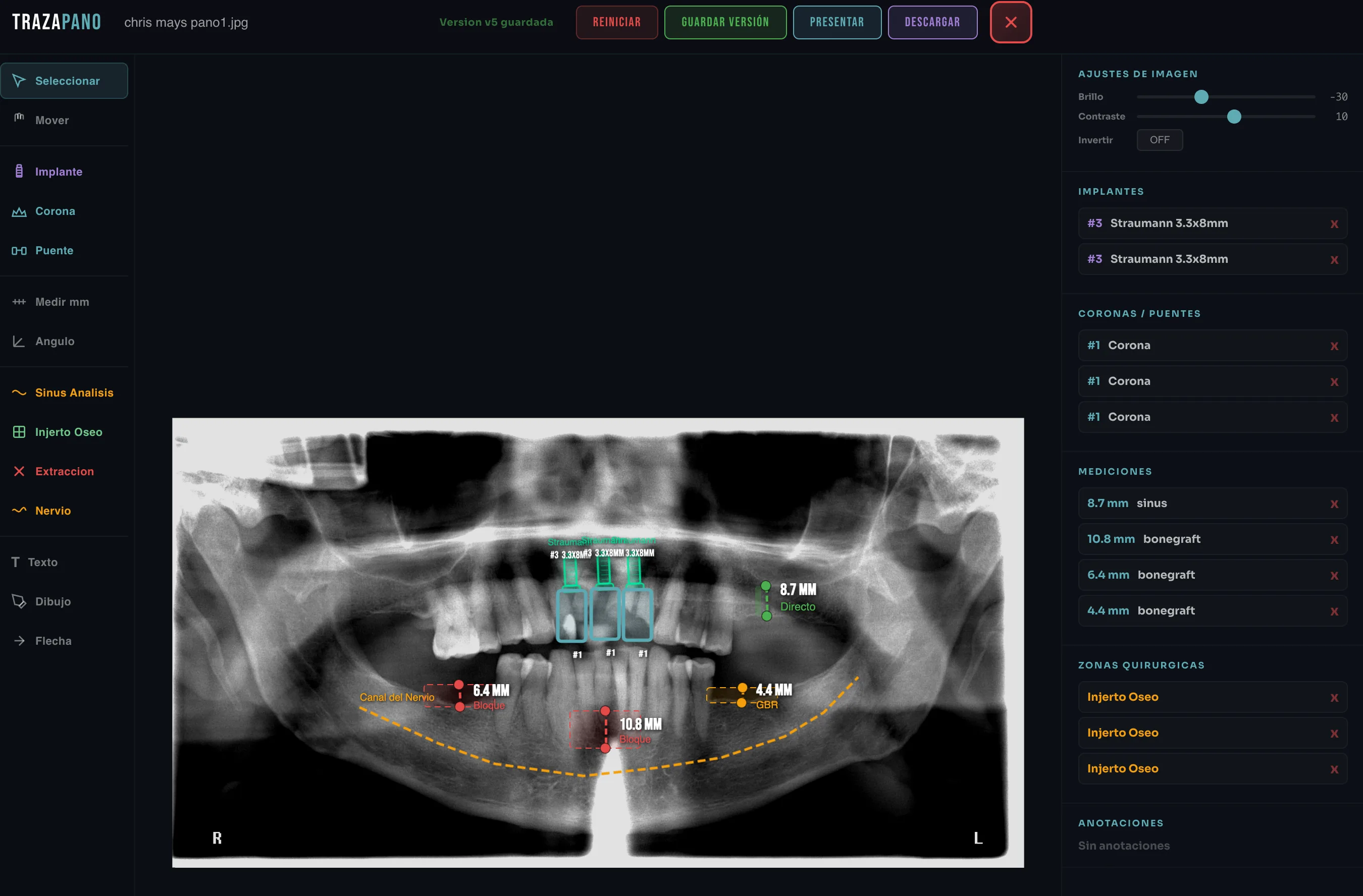
TRAZAPANO

Your panoramic talks. Surgical planning on panoramic X-rays.


No more drawing on printed X-rays with post-its. TrazaPano turns any panoramic into a surgical planning canvas with AI — real measurements, virtual implants, and automated 6-pass analysis.

TrazaPano — Surgical planning interface on a dental panoramic X-ray with Straumann implants, maxillary sinus measurements, nerve canal, and bone graft zones

Surgical planning on panoramic X-rays is still analog, imprecise, and disposable.

Drawings on paper

Surgeons print the panoramic and draw on it with markers. The plan lives on paper that wrinkles, stains, and gets lost.

No calibrated measurements

Without calibration, there is no way to know real bone height or available width. It gets eyeballed, and that creates surprises in surgery.

Ambiguous communication

The lab gets blurry photos of the hand-drawn plan and guesses the surgeon’s intent. Without digital data, communication breaks.

Plans that get lost

No versioning, no history. If the paper is lost, the plan is lost. Months of surgical analysis vanish in a drawer.

6-PASS AI VISION

GPT-4o analyzes your panoramic in 6 dimensions.

Each pass extracts a layer of clinical information that would take a human 45 minutes to document. AI does it in seconds.

1

Dental Spatial Map

Identifies each tooth with FDI coordinates and relative position on the panoramic.

#18 present, #17 present, #16 missing #15 present (mesial tilt 8deg) #14 root remnant Total: 24 teeth detected
2

Inventory & Quality

Catalogs teeth, restorations, root morphology, and image quality.

Restorations: 7 (3 amalgam, 4 composite) Endodontics: #36, #46 Existing implants: #26 (screw visible) Image quality: 8.2/10
3

Pathology Detection

Detects 25+ pathology types: caries, periapical lesions, fractures, resorption.

CARIES: #37 mesial (87% probability) PERIAPICAL: #46 radiolucency 4mm RESORPTION: #31 external cervical FRACTURE: #25 suspicious vertical line
4

Bone & Periodontal Analysis

AAP/EFP 2017 classification, Misch D1-D4 density, Klemetti index.

Periodontitis: Stage III Grade B Bone defect: #36 vertical 6mm Density zone #16: D3 (trabecular) Klemetti: C2 (eroded cortical)
5

Anatomical Structures

Maxillary sinuses, nerve canals, TMJ, third molars (Winter/Pell-Gregory).

R maxillary sinus: pneumatized (floor 3mm from #16) Inf. alveolar nerve: bilateral trace #48: Winter mesioangular, Pell-Gregory IIB TMJ: R condyle asymmetric
6

Implant Planning

Bone estimation, recommended implant size, augmentation needs, success probability.

Zone #16: bone 8mm height, 7mm width Recommended: 4.1x8mm with sinus lift Augmentation: lateral graft + membrane Success probability: 92%
IMPLANT PLACEMENT

Virtual implants with brand, diameter, and length.

Pick the implant system you use — Straumann, Zimmer, Implant Direct, Nobel, BioHorizons — and place virtual implants with real measurements on the panoramic. See the relationship with nerve and sinus before cutting.

  • Brand selector: Straumann BLX, BLT, Zimmer TSV, Implant Direct
  • Diameters: 3.3, 3.75, 4.1, 4.5, 5.0 mm
  • Lengths: 6, 8, 10, 12, 14 mm
  • Safety visualization with nerve and sinus
Straumann BLX Zimmer TSV Nobel Active
Size: 4.5 x 12mm
BLX 4.5x12
CALIBRATED MEASUREMENTS

Distance and angle in real millimeters.

Calibrate the panoramic with a known reference and measure bone heights, ridge widths, and inclination angles with submillimeter precision. Every measurement is saved with the plan.

  • Distance ruler (bone height and width in mm)
  • Angle measurement for implant inclination
  • Calibration tool with known reference
  • Real-time coordinate tracking
18.3mm 8.1mm 28°
BRIDGES & CROWNS

Prosthetic planning with FDI numbering.

Select abutments, pontics, single crowns, and extractions directly on the panoramic. The plan uses universal FDI numbering for unambiguous communication with the lab.

  • Single crowns with visual selection
  • Bridges with abutments + pontics
  • Extractions marked with visual X
  • Universal FDI numbering
UPPER ARCH — PLANNING
18
17
16
15
14
13
12
11
21
22
23
24
25
26
27
28
ABUTMENT: #14, #13, #24, #25 CROWN: #15, #23 EXTRACT: #16
BONE GRAFT ANALYSIS

Current bone level vs. target level.

Mark existing bone level and the level you need to reach with the graft. TrazaPano calculates the difference and clearly visualizes the augmentation zone needed at each surgical site.

  • Current vs. target bone visualization
  • Estimated graft volume calculation
  • Sinus lift planning
  • Documentation for the lab
ZONE #16 — GRAFT ANALYSIS
CURRENT
TARGET
DIFFERENCE
+6.2mm
Bone graft + membrane required
Current bone: 5.8mm Target: 12.0mm
Lateral sinus lift recommended before placement
NERVE IDENTIFICATION

Inferior alveolar nerve traced with precision.

Trace the path of the inferior alveolar nerve directly on the panoramic. See the safety distance between the planned implant apex and the nerve canal to avoid paresthesia.

  • Bilateral mandibular canal tracing
  • Implant-to-nerve distance in mm
  • Visual alert for risk zone
  • Mental foramen identified
FORAMEN FORAMEN 4.2mm SAFE DISTANCE
VIEWING MODES

Two modes. Two contexts.

Presentation Mode to explain the plan to the patient chairside. Surgery Mode to operate with a minimalist interface.

Presentation Mode
Implant #16 Treatment Plan — Patient: Martinez, J.

Presentation Mode

Clean interface with no toolbars. Just the plan and annotations. Perfect for explaining treatment to the patient in fullscreen.

Fullscreen for chairside
Surgery Mode
4.5x12 V Z R

Surgery Mode

Minimalist interface with high contrast for operating use. Only keyboard shortcuts and essential information visible during the procedure.

Minimalist for the OR
TECHNOLOGY

Built on battle-tested tools.

Every component is chosen for performance, precision, and reliability in a clinical context.

01 GRAPHICS ENGINE
Fabric.js Canvas
The same graphics engine used by Google, Microsoft, and Adobe for their visual editors. High-performance HTML5 canvas for object manipulation, layers, and real-time transforms.
CANVAS LAYER STACK
02 ARTIFICIAL INTELLIGENCE
6-Pass AI Vision
GPT-4o Vision analyzes your panoramic in 6 independent dimensions: dental map, inventory, pathologies, bone, anatomy, and implants. Each pass is tuned for a type of clinical finding.
1
OK
2
OK
3
...
4
...
5
QUEUE
6
QUEUE
03 CLINICAL FORMAT
Native DICOM Support
Direct loading of digital X-rays in DICOM format without external converters. Also supports JPG, PNG, and WebP for scanned or photographed panoramic X-rays.
panoramic_2026.dcm
DICOM 3.0 · 24.8 MB
OK
pano_scanned.jpg
JPEG · 3.2 MB
OK
x-ray.webp
WebP · 1.8 MB
OK
04 PERSISTENCE
Auto-save every 30 seconds
Never lose a surgical plan. TrazaPano auto-saves every 30 seconds with version history. If you close the tab or lose connection, your work is safe.
30s
Auto-save active
05 PROFESSIONAL WORKFLOW
Keyboard Shortcuts
V to pan, Z to zoom, R for ruler, A for arrow, T for text, I to invert. Professional flow without lifting your hands from the keyboard. Ctrl+Z to undo, Ctrl+Y to redo.
VPan
ZZoom
RRuler
AArrow
TText
IInvert
06 COMMUNICATION
Export Plan PDF
Generate a professional PDF with the annotated panoramic, measurements, planned implants, and AI findings. Email it to the patient, the lab, or file it in the chart.
surgical_plan.pdf

From paper to digital precision.

What changes when your surgical planning goes digital.

Before — Without TrazaPano
Printed panoramics with marker drawings
Eyeballed measurements without calibration
Paper plans that get lost or damaged
No automated pathology analysis
Ambiguous communication with the lab
No history or plan versioning
After — With TrazaPano
Digital canvas with calibrated tools
Real mm measurements with calibration
Versioned plans with 30s auto-save
AI analyzes 25+ pathologies in 6 passes
Exportable PDF with precise data for the lab
Full history with unlimited undo/redo

Your next panoramic,

planned.

With surgical precision.

14 days free. No credit card. Upload your first panoramic in 2 minutes and see what the AI finds.